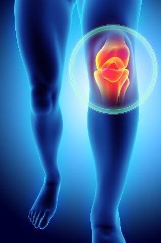
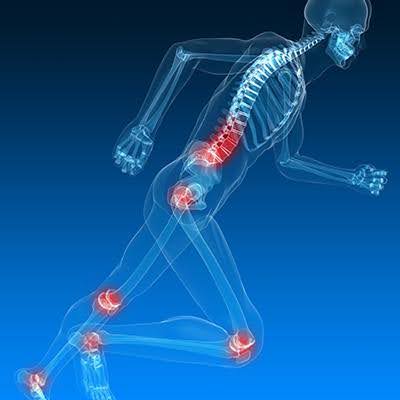
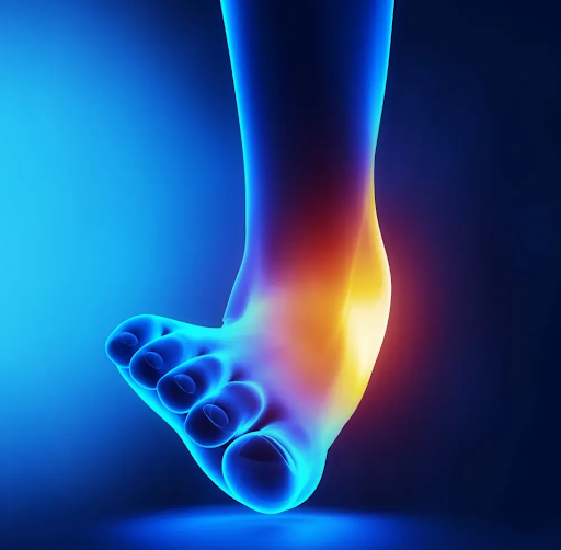
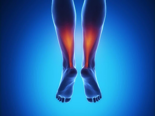
Sports Injuries

Sports Injuries we provide support for:

Indications

– Fractures with significant displacement or angulation
– Joint dislocations (e.g., shoulder, hip, elbow)
– Inability to perform reduction under local anaesthesia
– Paediatric fractures where cooperation is difficult

Procedure Overview

– General anaesthesia is administered with airway management (e.g., endotracheal tube or laryngeal mask airway).
– The surgeon applies specific techniques to realign the bone or joint.
– Fluoroscopy or X-rays may be used to confirm alignment.
– Immobilization with a cast, splint, or brace.
– Post-reduction X-ray ensures proper alignment.
– Pain management and monitoring for complications.

Risks

– Failure to achieve reduction
– Re-displacement or instability
– Nerve or vascular injury
– Complications related to anesthesia

Follow-up

– Serial x-rays imaging and clinical assessments to ensure healing.
– Physiotherapy may be required to restore function.

Indications for Internal Fixation:

– Displaced fractures (misaligned bone fragments).
– Open fractures (where the bone breaks through the skin).
– Comminuted fractures (multiple bone fragments).
– Fractures that fail to heal properly with non-surgical methods.

Types of Internal Fixation Devices:

– Plates and Screws – Metal plates are attached to the bone with screws to hold fractured fragments in place.
– Intramedullary Nails/Rods – Long metal rods inserted into the bone’s hollow center, commonly used for long bone fractures like the femur or tibia.
– Screws – Used alone or with other devices to hold bones together.
– Wires and Pins (Kirschner wires or K-wires) – Often used for small bone fractures or temporary fixation.

Benefits

– Provides strong and stable fixation.
– Enables early mobilization and faster recovery.
– Reduces the risk of malunion or non-union.

Risks

– Infection 
– Implant failure or loosening.
– Delayed or non-union of bones.
– Nerve or blood vessel damage.
– After surgery, rehabilitation with physiotherapy is essential for full recovery.

Indications


– Severe fractures (open, unstable, or comminuted fractures)
– Complex limb injuries (such as fractures with soft tissue damage)
– Limb lengthening
– Correction of bone deformities
– Infected fractures (osteomyelitis cases)
– Temporary stabilization before definitive surgery

Types of External Fixators

– Unilateral fixators – Simple bars on one side of the limb.
– Circular fixators– Used for deformity correction and limb lengthening.
– Hybrid fixators – Combination of both types for complex cases.

Benefits

– Minimally invasive (avoids large incisions)
– Allows soft tissues to heal while stabilizing bones
– Can be adjusted without additional surgery

Risks

– Risk of pin-site infections
– Can be uncomfortable for the patient
– Requires careful maintenance and follow-ups

Causes of Non-Union

– Poor blood supply to the bone
– Inadequate stabilization (movement at the fracture site)
– Infection at the fracture site
– Smoking or poor nutrition, which affect bone healing
– Medical conditions like diabetes or osteoporosis

Symptoms of Non-Union

– Persistent pain at the fracture site
– Swelling or tenderness that doesn’t go away
– No signs of healing on X-rays over months
– Weakness or inability to use the affected limb

Treatment Options

– Surgery: May involve bone grafting, internal fixation (plates, screws, rods), or external fixation to stabilize the bone
– Bone stimulation: Electrical or ultrasound stimulation can encourage healing
– Medications: Certain drugs (like bone growth factors, BMPs, Bone grafts) may help
– Lifestyle changes: Stopping smoking, improving diet, and managing underlying conditions